- Visibility 147 Views
- Downloads 10 Downloads
- DOI 10.18231/j.ijpi.2024.032
-
CrossMark

Exploring vestibuloplasty techniques- An insight from 3 cases
- Author Details:
-
 Shruthi Reghunath *
-
Aswathy Jayasree
-
Bilha Joy
-
Roshni Ramesh
-
Raseena Beevi Nafeesa
-
Abdurasheed Edakkot Mathamkuth
Introduction
Periodontal plastic surgery procedures aim to address various issues affecting the gums, alveolar mucosa, and bone, such as defects arising from anatomy, development, trauma, or plaque-related diseases. These procedures not only serve aesthetic purposes but also contribute to maintaining optimal oral hygiene. The oral vestibule refers to the cavity within the mouth, bordered medially by the teeth and gingiva, laterally by the cheek and lip mucosa, and apically by movable and immovable mucosal borders. Vestibular depth is measured either from crest of lip or from coronal border of the attached gingiva to depth of mucobuccal fold.[1] Adequate vestibular depth is crucial for ensuring proper oral hygiene, as a shallow vestibule can lead to food accumulation and hinder the maintenance of oral cleanliness. Additionally, when combined with inadequate attached gingiva, shallow vestibules may result in marginal tissue pulling and gingival recession, exacerbating plaque accumulation, gingival inflammation, tooth mobility, bone loss, and further gingival recession. Furthermore, an appropriate vestibular depth is essential for the retention and stability of removable dentures. [2]
Vestibuloplasty, a surgical procedure, aims to deepen the oral vestibule by altering soft tissue attachments. Several techniques are employed to deepen shallow vestibules by modifying soft tissue attachments. These include submucosal vestibuloplasty, secondary epithelial vestibuloplasty, and soft tissue graft vestibuloplasty. Periodontal procedures in the new millennium have moved on from being extensively aggressive to minimally invasive. Recent advancements in intraoral surgical techniques include use of lasers with the advantages like greater precision and a relatively bloodless surgical field. In this case report, we compare three different vestibuloplasty techniques: conventional Clarks surgical vestibuloplasty, vestibuloplasty using diode lasers, and a modified open submucosal vestibuloplasty without sutures. Each technique offers unique advantages and considerations, contributing to the growing body of knowledge in the field of periodontal plastic surgery.
Case Presentation
Case 1
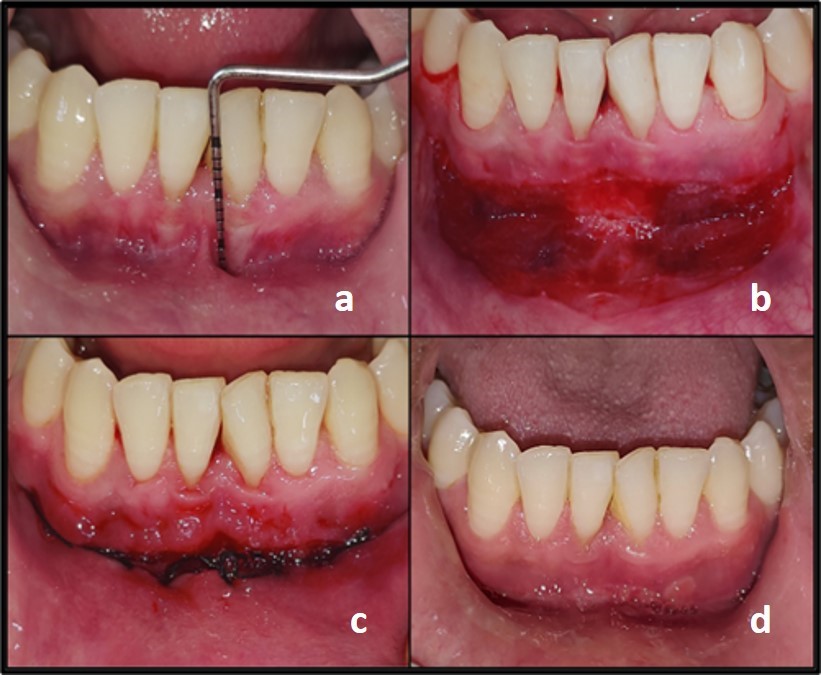
A 24-year-old male presented to the Department of Periodontics and Implantology with the chief complaint of difficulty in brushing teeth on the lower front teeth region for the past eight months, also complained of rapid accumulation of deposits over that area. On clinical examination, the patient had gingival recession in relation to lower incisors and reduced vestibular depth in the lower anterior area. After two weeks of non-surgical periodontal therapy, vestibuloplasty (Clark’s technique) was performed ([Figure 1]). Under local anaesthesia, horizontal incision using No.15 blade was placed at mucogingival junction from lower left to right canine which was followed by supraperiosteal dissection up to the desired vestibular depth. The mucosa on the inner side of the lip was then undermined and the mucosal flap was secured to the periosteum, deep into the sulcus, with non- resorbable 5-0 suture at the depth of vestibule. The raw area over the alveolar bone was left to heal by secondary intention. Coe pack periodontal dressing was placed for wound protection.
Case 2
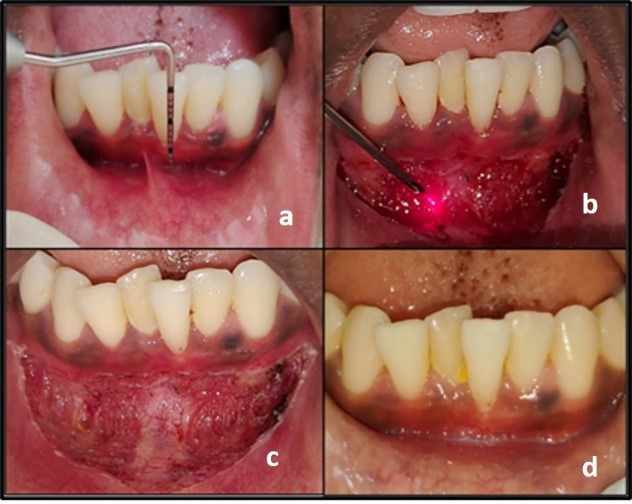
A 21-year-old female presented to the Department of Periodontics and Implantology, reporting of bleeding from her lower front teeth region for the past 1 year. The patient was having non-contributing medical history. Upon examination, visible plaque and calculus deposits were observed along with inflamed marginal gingiva and interdental papilla in the mandibular anterior region. Positive bleeding on probing was noted. Overall oral hygiene was satisfactory except for the lower anterior labial region, where inadequate vestibular depth and a positive frenal pull were evident around lower left canine to right canine. A vestibuloplasty procedure was performed utilizing a diode laser with a wavelength of 810 nm. The soft tissue laser was operated in surgical mode at a power of 3 watts ([Figure 2]). An initial incision was made along the mucogingival junction from the right lower canine to the left lower canine. Subsequently, all muscle fibers were meticulously dissected, including the labial frenum using laser, and the vestibule was deepened to achieve the desired depth. Saline irrigation was performed, and a coe-pack dressing was applied along with detailed post-operative instructions.
Case 3
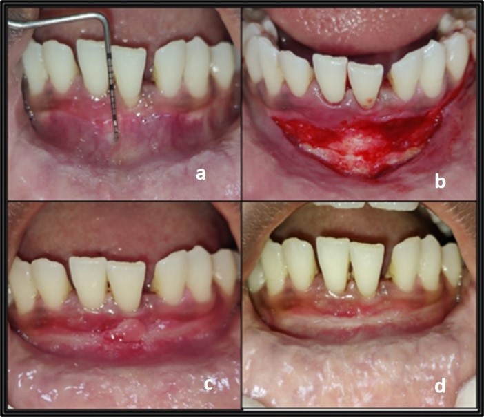
A 29-year-old female presented to the Department of Periodontics and Implantology, reporting of bleeding from her lower front teeth and gum recession. She also mentioned difficulty in maintaining regular oral hygiene, hindering toothbrush movement. The patient did not have prior dental history. Intraoral examination showed a shallow vestibule and spacing between lower front teeth, with a positive tension test on the lower front teeth. Following phase I therapy, a modification of open submucosal vestibuloplasty was planned to address the shallow vestibule. For vestibuloplasty, a horizontal incision was made at the mucogingival junction from lower left canine to right canine followed by supraperiosteal dissection to achieve the desired vestibular depth. The labial mucosa was then undermined, and periosteal scoring was performed using blade no. 11. The incision was left unsutured, and a periodontal pack was applied deep into the vestibule after ensuring adequate hemostasis ([Figure 3]).
Post-surgical instructions were provided for all the cases, and the patients were prescribed antibiotics (Cap Amoxycillin 500 mg three times a day for 5 days) and analgesics (Tab Paracetamol 500 mg two times a day for 3 days). Tooth brushing was restricted at the surgical site for the first week, with extraoral cold compression recommended for the first day. All the patients were reviewed on day 1,3 and 7 and examined for pain using visual analogue scale and clinical outcomes were assessed on day 7 and day 21.
Results
An increase in vestibular depth was attained by all the three procedures. In the traditional Clarks technique described in case 1, significant gain in vestibular depth was observed during the postoperative reviews at day 7 and day 21. Patient experienced mild pain and swelling in the first two days after surgery, with relief starting on the third day. Healing progressed smoothly, without scar tissue formation, and there was a substantial gain in vestibular depth. Patients pain perception was measured using visual analogue scale (VAS) on day 1, 3 and 7. When the diode laser was used, patients experienced notable swelling and pain, as well as difficulty eating, for the first 5 days. However, these symptoms improved thereafter. Although the 7-day postoperative review showed a good increase in vestibular depth without scar tissue formation, there was a slight reduction in the gained depth when reviewed at 21 days. The modified open submucosal vestibuloplasty, which did not involve sutures, achieved a vestibular depth gain comparable to the conventional Clarks technique. However, scar tissue formation was observed in the lower incisor areas at the 7-day postoperative review ([Figure 3]c). Patients were advised to apply an astringent locally, and by the 3rd -week, patients reported symptomatic improvement and resolution of scar tissue. All patients experienced pain, swelling, and difficulty eating for the first 5 days postoperatively. The VAS score for all techniques obtained on day 1,3, and 7 is enumerated in [Table 1].
|
VAS score |
Clark’s technique |
Lasers |
Modified open submucosal vestibuloplasty |
|
Day 1 |
6 |
7 |
7 |
|
Day 3 |
4 |
6 |
5 |
|
Day 7 |
2 |
3 |
3 |
Discussion
Vestibuloplasty, also known as sulculoplasty, is a surgical intervention that aims to alter the relationship between the gingiva and mucous membrane. The procedure can involve deepening the vestibule, repositioning the frenulum or muscle attachments, and expanding the zone of attached gingiva. [3] It is used to address issues such as preventing further gingival recession, restoring attached gingiva, enhancing vestibular depth, improving plaque control, and boosting resistance to masticatory trauma. Additionally, vestibuloplasty can be performed for cosmetic purposes and to create a better base for dentures in edentulous patients, thereby improving retention and stability. In cases where tension from the frenulum affects tissue around implants, leading to inflammation and recession, vestibuloplasty can help alleviate the issue.
There are some contraindications to consider before proceeding with vestibuloplasty, such as significant bone loss after traumatic tooth extraction, notable bone resorption due to periodontitis, alveolar bone atrophy following tooth removal, and patients with severe systemic illnesses. Careful assessment of these factors is crucial to ensure successful outcomes and minimize risks. Vestibuloplasty techniques are categorized into three main types: mucosal advancement vestibuloplasty, secondary epithelization vestibuloplasty, and grafting vestibuloplasty. [4] [Table 2] outlines the various techniques of vestibuloplasty.
|
Technique |
Sub-classification |
|
1.Mucosal advancement vestibuloplasty |
a. Closed submucosal vestibuloplasty b. Open submucosal vestibuloplasty |
|
2. Secondary epithelialization (Reepithelialization vestibuloplasty) |
a. Kazanjian’s technique b. Clark’s technique c. Lipswitch technique |
|
3. Grafting vestibuloplasty |
a. Mucosal grafts b. Skin grafts |
|
4. Others |
a. Laser vestibuloplasty b. Electrocautery |
Clark's technique, introduced by Clark in 1953, is a secondary epithelialization vestibuloplasty method used in cases where there is adequate bone, but the mucosa is either insufficient or of poor quality. The technique is based on the principle that exposed connective tissue surfaces contract, while those covered with epithelium contract less, and exposed bone surfaces do not contract. Proper undermining and fixation of the epithelial flap are necessary to prevent soft tissues from reverting to their original position, which requires some overcorrection. The conventional Clark's technique has been effective, resulting in a significant increase in vestibular depth with minimal wound contracture and relapse. However, the surgical process is complex, time-consuming, and requires a high level of expertise.
In contrast, laser surgery provides several benefits, including precise incisions, a bloodless field, less requirement of local anaesthesia, and shorter procedure time. [5] It also creates an aseptic environment and eliminates the need for sutures. Despite these advantages, patient reported more postoperative discomfort, and there was a slight reduction in gained depth by the 21st day post-surgery. The high cost of laser equipment and potential hazards associated with its use raise questions about its routine application in oral surgical procedures.
In the third case, a modified open submucosal vestibuloplasty was performed without the use of sutures. This approach, based on the technique advocated by Wallenius, focused on mucosal advancement and was selected due to the presence of adequate bone and healthy mucosa. The procedure offered a simplified alternative to traditional methods, resulting in a shorter operative time and outcomes similar to Clark's technique. However, the lack of sutures presented challenges in achieving hemostasis. Additionally, healing was not optimal, as evidenced by the presence of scar tissue one week postoperatively. Further refinement of the technique may be needed to address these concerns and improve patient outcomes.
In a recent study conducted by Karas et al. (2023), the outcomes of diode laser and conventional surgery were compared in terms of vestibular depth gain. The study revealed that conventional surgery resulted in a higher vestibular depth gain compared to diode laser surgery. Furthermore, low-level laser therapy (LLT) did not demonstrate any significant benefit in promoting mucosal wound healing. [6] Kalakonda et al. assessed patient perceptions of pain and healing outcomes following mandibular vestibuloplasty, comparing scalpel and laser techniques. Patients were evaluated on the 1st, 3rd, and 7th days post-surgery. The study found that patients treated with laser reported lower pain and discomfort, as measured by visual analog scale (VAS) scores, compared to those who underwent the procedure with a scalpel. Additionally, the laser group exhibited improved healing outcomes. [7]
Rogoleva et al. (2022) reported similar findings, showing that laser treatment resulted in better healing outcomes and fewer complications, such as pain and swelling, post-operatively. [8] Bhullar et al. compared pain perception following vestibular extension procedures using three different techniques: scalpel, laser, and cautery. The study supported the use of electrocautery in soft tissue procedures, such as vestibular depth extension, as it provided better patient perceptions of pain and discomfort than scalpel and laser techniques. [9] A retrospective study by Neckel compared vestibuloplasty outcomes using laser and scalpel techniques, finding no significant difference in vestibular height gain between the two methods. However, patient perceptions slightly favoured laser treatment over scalpel. [10] Lastly, a study by Abdelfattah et al. highlighted the advantages of Er:YAG laser vestibuloplasty over conventional surgery, noting better patient perceptions and healing outcomes. [11] Although most studies suggest that laser techniques offer better patient pain perception and improved surgical outcomes, our findings indicates that Clark's technique produced better outcomes. Furthermore, both techniques demonstrated similar levels of pain perception.
Conclusion
This case report explores three distinct methods for increasing vestibular depth: the traditional Clarks technique, the use of lasers, and a modified open submucosal vestibuloplasty. Each method has its own set of advantages and disadvantages. The greatest increase in vestibular depth was achieved with the traditional Clarks technique. Patients reported similar levels of pain across all three techniques. The modified open submucosal vestibuloplasty is a relatively straightforward and time-efficient procedure, providing results comparable to those of the Clarks technique. Even though there was scar tissue formation in the early stages of healing, complete resolution of the scar with adequate healing was observed in subsequent visit. The laser approach offers several benefits, including hemostasis, precision, and the elimination of the need for sutures. Each technique for vestibuloplasty presents a unique set of advantages and disadvantages and the choice of technique depends on individual patient needs, surgical goals, and available resources. When compared to other studies our findings showed better vestibular depth gain with traditional Clark’s technique and comparable pain score with all three methods.
Limitations
In this case report we have compared only one case in each category which provide only limited evidences. Further comparative studies with large sample sizes are required to gather more substantial evidence on each technique's efficacy and safety.
Conflict of Interest
The authors declare that there is no conflict of interest.
Source of Funding
None.
References
- V Ward. A technique of measurement of the depth of the vestibular fornix in the mandibular anterior region. J Periodontol 1976. [Google Scholar]
- C Ochsenbein. Newer concepts of mucogingival surgery. J Periodontol 1960. [Google Scholar]
- . American Academy of Periodontology (AAP). Glossary of periodontal terms [internet]. 2001. [Google Scholar]
- T Froschl, A Kerscher. The optimal vestibuloplasty in preprosthetic surgery of the mandible. J Craniomaxillofac Surg 1997. [Google Scholar]
- S Coleton. Lasers in surgical periodontics and oral medicine. Dent Clin North Am 2004. [Google Scholar]
- M Karas, S Gunpinar. The use of low level laser therapy in conjunction with diode laser-assisted and conventional vestibuloplasty: Comparison of wound healing and vestibular depth gain. J Stomatol Oral Maxillofac Surg 2023. [Google Scholar] [Crossref]
- B Kalakonda, S Farista, P Koppolu, K Baroudi, U Uppada, A Mishra. Evaluation of Patient Perceptions After Vestibuloplasty Procedure: A Comparison of Diode Laser and Scalpel Techniques. J Clin Diagn Res 2016. [Google Scholar]
- SR Gjurovski, T Piperevaliev, VT Stojmenova, B Nikolovski. Bruno (2022) Conventional and laser vestibuloplasty comparison. In: 21 Kongres stomatologa Srbije, 20-23 Oct 2022. 2022. [Google Scholar]
- S K Bhullar, V Goel, A Bhullar, L Goyal, V Mehta, T Nanda. Comparative evaluation of pain in vestibular depth extension procedure using scalpel, electrocautery and diode laser. J Dent Spec 2017. [Google Scholar]
- C Neckel. Vestibuloplasty: a retrospective study on conventional and laser operation techniques. PROC. SPIE 3593. Lasers Dent . [Google Scholar]
- MY Abdelfattah, H Moharam, M Fahmi, A Kossar. Assessment of Patient Perception, Healing Outcomes and Gained Vestibular Depth of Er: Yag Laser Versus Conventional Surgical Mandibular Vestibuloplasty. Int J Dent Sci Res 2015. [Google Scholar]