- Visibility 154 Views
- Downloads 31 Downloads
- DOI 10.18231/j.ijpi.2024.024
-
CrossMark

Ultrasound in periodontology and implantology
Introduction
The term ultrasound refers to sound waves with frequencies higher than those of audible sound, which is typically above 20,000 hertz according to Laird WR and Walmsley AD (1991).[1] Pierre and Jacques Curie (1880) identified that when crystals of various substances are subjected to mechanical strains, their surfaces acquire electrical charges. As a natural byproduct, it was soon discovered that crystals and metals treated in this way oscillated at a particular frequency and amplitude when exposed to an alternating electrical field of the right size and composition. "Physical and Biologic Effects of High Frequency Sound Waves of Great Intensity" was published by Wood and Loomis in 1927.[2] Ultrasound has mainly been utilized in the medical field for treating neuromuscular and musculoskeletal conditions since then.[2] The initial application of the magnetostrictive cutting device in industry involved creating cavities in synthetic sapphires to accommodate gold inserts. The first instance of employing this cutting technique in dentistry was documented by Matthew C. Catuna in 1953.[3] In the past, these tools were utilized alongside abrasive slurry to prepare tooth cavities before restoration. The introduction of high-speed drills led to a shift in technology towards ultrasonics and power scaling in the early 1960s, completely transforming mechanical debridement.
In 1955, Zinner[4] introduced ultrasound to periodontal procedures with ultrasonic scalers, which have seen numerous modifications since then. As a result, compact devices have replaced the original large, heavy units. A range of site-specific, smaller tips—some of which have been dubbed microultrasonic which have replaced the single, clunky universal tip. Johnson and Wilson [5]found that using ultrasonic tips instead of traditional scaling methods allowed for the appropriate removal of calculus more quickly. They also came to the conclusion that scaling with ultrasonic requires just little pressure. Very little bleeding is related to ultrasonic therapy, cementum under calculus is mostly unaffected, and patient response to the device has been positive, particularly in ANUG patients. There was curiosity in the nature of the cleaning process in the late 1980s and early 1990s when it was demonstrated that acoustic microstreaming and cavitation play different roles (Walmsley AD 1982). [6], [5]
Basic principle
A subfield of acoustics called ultrasonics studies sound waves at frequencies higher than human hearing. Using high frequency sound waves, ultrasound imaging, often known as sonography or ultrasound scanning, is a technique for creating images from inside the human body. Ultrasonic beams are attenuated by a mixture of absorption, reflection, refraction, and diffusion when they travel through or interact with tissues with varying acoustic impedence of one another. The echoes of the sound waves are captured and shown as a visual representation in real time. Acoustic energy is used in both the transmission and reflection of ultrasound. The transducer, a device that transforms electrical energy into acoustic energy, both propagates and receives the reflection of a pulse. Transducers are devices that transform electrical energy into ultrasonic waves for use in medicine.
Usually, piezoelectricity or magnetostriction are used to accomplish this. When exposed to a magnetic field, magnetostrictive devices experience physical dimension changes. Usually, a ferromagnetic stack is placed within a solenoid that receives a direct current in order to do this. This causes tensions that cause the material to shift shape. The stack will then vary shape at twice the frequency of the applied magnetic field when an alternating current is run through the solenoid. Because it is a reliable and simple to construct technology, magnetostriction with a laminated ferromagnetic stack is frequently employed in the design of ultrasonic scaling instruments. Magnetostrictive instruments use a metal rod attached to a scaling tip or a stack of flat metal strips operating between 18,000 and 45,000 cps. A magnetic field is produced around the stack or rod transducer in the handpiece when an electrical current is applied to a wire coil, causing it to contract. The tip then vibrates as a result of an alternating current creating an alternating magnetic field. Magnetostrictive units can travel in a nearly linear, elliptical, or circular manner at the tip, depending on the type of unit and the length and form of the tip. With magnetostrictive tip movement, all tip surfaces can be activated concurrently, giving the user the choice to adapt the tip to the tooth surface by using its front, rear, or side. The foundation of a piezoelectric system is the observation that in the presence of an electrical field, some crystalline structures, like quartz, will undergo shape changes. A piezoelectric crystal will exhibit an oscillating shape change at the applied frequency if an alternating voltage at an ultrasonic frequency is placed across it. After that, this is transferred to the functioning tip.
Lead zirconate titanate is currently the most commonly used piezoelectric material (PZT). While some have been developed for use in dentistry, piezoelectric generators are more efficient at frequencies in the MHz region as opposed to the KHz range. Nevertheless, these instruments are more delicate than their magnetostrictive counterparts due to the weak shock resistance of the crystalline structure. The crystals inside the handpiece undergo dimensional changes when electricity is delivered over their surface, activating the piezoelectric device, which operates in the 25,000–50,000 cps range. The resulting vibration causes mostly linear tip movement, typically allowing just two sides of the tip to be active at any given time. The majority of modern ultrasonic technology has developed to the point where computer chips are used to control the continuous power to the tip (Pattison AM 2006). [7]
Properties of ultrasound
Ultrasound waves do not pass through air.
Ultrasound can only visualize the exterior of bony structures and not their interiors because it has difficulty in penetrating bone
In contrast to X-rays, which create images by transmitted radiation, ultrasonography creates images through the reflected component of the beam.
The same principles that underpin ship sonar, bat sonar, and angler fish detectors also underpin ultrasound imaging. Echoes that are created when sound travels through the body can be utilized to determine an object's size, shape, consistency (fluid, solid, or mixed), and distance from the source.
A noninvasive, reasonably priced method for real-time imaging of superficial tissues is ultrasound. (Nordic Frederiksen, 2004) [8]
Ultrasonic imaging involves no ionizing radiation
In water, ultrasound waves travel at a virtually constant speed of about 1500 m/s. The sound wave velocity in soft tissue is comparable to that in water.
Basic effects of ultrasound
Thermal effects
Tissue temperature rises as ultrasonic waves travel through tissues because their energy is absorbed and released as heat. The amount of the temperature spike, the duration of the temperature rise, and the tissue's thermal sensitivity all affect the tissues. Most tissues will naturally respond physiologically by varying the blood flow in the area as a result of the arterioles' reflexive relaxation. A temperature rise of less than 10ºC will only cause a slight overall increase in local metabolic rate. The increased blood flow that follows will tend to moderate heating effects within a limited temperature increase. Nonetheless, tissue injury is an inevitable consequence of exceedingly high temperatures (Laird WR, Walmsley AD 1991). [1], [8]
Cavitation
When it comes to ultrasonic, cavitational activity refers to a continuous range of bubble activity in a liquid medium. It can vary from severe and destructive behavior of vapor-filled cavities (transient cavitation) in high amplitude sound fields to calm linear pulsation of gas-filled bodies in low amplitude sound fields (Williams AR. 1983).[9] The energy produced by these bubbles has the potential to cause shock waves or hydrodynamic shear fields that could damage biological tissues. It is the creation of these powerful disruptive forces that helps remove calculus and plaque during ultrasonic scaling. Cavitation cannot occur unless there are gaseous particles, also known as cavitation nuclei, present in the medium. When there is an ultrasonic field present, a bubble will expand and pulse in response to the pressure oscillations the field creates. Transverse waves are created on the bubble's surface as it pulses, and as the ultrasonic amplitude rises, these waves become deformed and unstable. Around the original bubble, microbubbles will form and serve as fresh locations for cavitational action. Microbubble formation coincides with the start of transitory cavitation, in which the bubbles exhibit a collapse.
A phenomenon when the gas inside the bubble reaches temperatures of thousands of degrees Celsius and pressures of several thousand atmospheres. The shock waves released during the last phases of bubble collapse or the high-velocity liquid jets produced by the nonlinear motions of the bubble face are what cause the demanding consequences of transitory cavitation. Micronuclei can easily form at low ultrasonic frequencies, such as 20.40 KHz, and this can lead to temporary cavitation. Human blood cavitation can lead to platelet and erythrocyte lysis as well as a thrombogenic effect (Walmsley AD, 1991). [1] This could account for the decreased bleeding that occurs after surgery with ultrasonic tools.
Acoustic microstreaming
A complicated steady state streaming pattern forms in the liquid near the bubble surface as a result of the fast cyclical volume pulsation of a gas bubble (Walmsley AD. 1991).[1] A phenomenon known as "acoustic microstreaming" occurs in fluid environments like water and is typified by the generation of strong shear stresses. It can be shown around a stationary cylinder inside an oscillating fluid or an oscillating solid cylinder inside a fluid. [1]
The acoustic microstreaming that surrounds ultrasonic scalers is contingent upon the amplitude of displacement, tip direction, and the existence of a water medium. It depends on the distance from the oscillating tip, the tip geometry, and the tip orientation, but it rises with increasing displacement amplitude. The patterns' dimensions show how quickly the streaming velocity changes with distance. As a result, even though the velocities are only a few centimeters per second, the gradients caused by the velocity change rate will create significant hydrodynamic shear stresses near the oscillating object—the probe or gas bubble—which could disrupt or harm biological tissues or cells. Periodontal disease-related subgingival biofilm breakdown may be facilitated by acoustic microstreaming.
Blood flow and cells, including human platelets, exposed to probes operating at 20 kHz (the frequency used in dentistry) may also be disrupted by acoustic microstreaming. Higher amplitudes may cause blood artery obstruction due to the formation of emboli by gelatinous platelet aggregates (Khambay BS, Walmsley AD. 1999). [10]
Chemical effects (sonochemicals)
When teeth were scaled, sonochemical compounds were also formed in addition to the mechanical cavitational effects. Ions in the propagating medium are released rapidly and intensely upon the agitation of ultrasonic vibrations. Ultrasonic cavitation, which is akin to ionizing radiation, interacts with specific chemicals or gases in aqueous solutions, such as dissolved air, oxygen, and nitrogen, by means of free radicals generated from the breakdown of water molecules. Considering their chemical activity, free radicals and other chemicals generated within the solution (H2O2, nitrous, and nitric acids) are particularly important to biology. The geometry of the scaling tip and the displacement amplitude are connected to the free radicals generated.
Radiation forces
An ultrasonic beam exerts a radiation force on any medium or item in its path, which tends to push the material in the direction of the propagating wave. Although this force is tiny, it can be amplified and act across a short distance in a standing wave field, driving dense particles in the medium to areas where the amplitude of the acoustic pressure is at its highest. This could result in localized blood cell aggregation in blood arteries, which would be stasis. Within a standing wave Field, cavitational activity may also be enhanced by radiation forces.(Walmsley AD, Laird WR 1991) [6]
Secondary effects of ultrasound
Responses that a tissue may create or evoke during ultrasonic irradiation are known as secondary effects of ultrasound.(SJ Evan, 1960) [11]
Direct application of frictional movement vibrations at a frequency of 25 KHz can cause coagulation in the tissue.
If the propagation medium is constantly flowing between the tool and the tissue, a light massage may cause a hyperemia without destroying any tissue.
When ultrasound is administered to tissues with a high fluid content, the tissue will produce bubbles or begin to degas (cavitation).
Tissue surgery can be performed using ultrasonic instruments of the right design and frequency to cut turgid tissue that is either frozen solid or fluid-filled.
Other tissue effects
Researchers have shown that myalgia and tendon extensibility have improved with the use of high frequency vibrations.
Research in medicine has demonstrated that using ultrasonic therapy can soften scar tissue, especially burn scar tissue. As a form of scar tissue, the fibrotic gingiva associated with persistent gingivitis underwent ultrasonic imaging and produced findings that are comparable to those reported.
Coagulating the surface of soft tissue by rubbing or pressing the tip of a vibrating tool against it results in a soft tissue curettage. This type of curettage can be applied to the buccal or labial sides of the gingiva, or it can be done inside the crevice.
Ultrasonic vibrations applied to experimental animals' gingiva cause tissue continuity to be disrupted, lifting off epithelium, severing collagen bundles, and changing the shape of fibroblast nuclei. Targeted towards tissue interfaces, namely the junction between connective tissue and epithelium, ultrasonic vibrations propagate laterally and push the epithelium off. The collagen bundles are forced apart mechanically and the connective tissue beneath becomes dehydrated. A type of coagulated wound is the deficiency in the tissues that is so generated.
Ultrasound is an additional tool for gingival surgery in addition to soft tissue curettage. Gingival tissue can be removed with periodontal curettes that have been ultrasonically vibrated and sharpened to a razor's edge.
The development of a non-invasive periodontal assessment tool with ultrasound technology holds great promise for providing real-time, high-yield information on clinical features like pocket depth, attachment level, tissue thickness, histological change, calculus, bone morphology, and tooth structure evaluation for fracture cracks.
The first time ultrasonography was used in periodontology was by Spranger in 1971. He attempted to measure the alveolar crest height in patients with periodontal disease. An ultrasonic transducer aimed perpendicular to the tooth's long axis in an attempt to scan the crest of alveolar bone. The measuring of gingival thickness with an ultrasonic device with a 5 MHz transducer. Despite the fact that these studies demonstrated the viability of ultrasonic imaging in dentistry, researchers also shown that the level of periodontal attachment can be deduced from the echoes that an ultrasonic instrument detects from the hard tissue of the tooth surface.
Discussion
Application of ultrasound in diagnosis
Periodontal ultrasonography
The non-destructive evaluation sciences laboratory at NASA Langley Research Centre regularly uses an ultrasound-based time-of-flight approach to evaluate material thickness and, in certain situations, length. This technique is the source of the ultrasound technology utilized in periodontal ultrasonography.
This technology has mostly been used in aircraft skin thickness measurements for corrosion detection and bolt length measures for bolt tension. An ultrasound probe functions similarly to a sonogram. A ultrasound records and shows an image of the unborn face by pressing a probe on the body, sending a laser through the womb and recording the echoes that are returned. The same technology is used to image periodontal structures, mostly through the use of a very small probe that transmits and receives ultrasonic signals. Computer software immediately filters out all echoes and creates an image of the attachment level, pocket depth, etc. (Farr C. 2000). [12]
Periodontal ultrasonic probe
It is made up of a transducer that is contained at the base of a hollow conical tip in a contraangled handpiece. This is in charge of both sound wave emission and reception. Acoustic beam is focused into periodontal tissue by the hollow tip. To provide an acoustically tapered interface with a throat area of approximately 0.5 mm, a transducer is installed at the base of a dual taper, convergent, divergent coupler. An active area reduction from transducer element to aperture is represented by a throat area of 1.5 mm. The gingival margin's tiny window and geometry both require this kind of reduction. The capacity of the ultrasonic probe to inspect the interdental area is an additional benefit of achieving a small tip size. A computer, monitor, keyboard, separate electron box for controlling the water pressure, and foot pedal are among the necessary equipment needed to operate the probe. Everything put on a sizable cart for easy mobility. To guarantee an effective coupling of ultrasonic energy to tissues, the probe tip integrates a tiny flow of water. Water can be drawn from a dental chair or an IV-style sterile bag that is hanging.
Working of ultrasonic probe
The ultrasonic probe tip is held vertically, parallel to the tooth's long axis. To ensure that all of the water has been completely coupled into the gingival sulcus, the tip is gently put on the gingival border until a minor blanching of the gingiva is seen. A foot pedal is then used to activate the probe. A tiny jet of water and a narrow ultrasonic wave beam enter the sulcus when the foot pedals are depressed. An ultrasonic probe listens for returning wave echoes after projecting a narrow frequency (1.20 MHz) ultrasonic pulse into the gingival sulcus or periodontal pocket. When an ultrasonic beam enters tissues, it can be scattered, reflected, or absorbed. The machine receives the reflected portion and uses it to recreate the ultrasonic image. A tiny sensor in the handpiece records the echoes of these sound waves as they bounce off periodontal tissues, and a computer connected to the ultrasonic device analyzes the data concurrently. A computer collects incoming data while the examiner moves the probe tip across the gingival edge. Artificial intelligence algorithms are then used to interpret the recorded data into estimates of the probing depth in millimeters.
The ultrasonic probe tip is softly placed on the gingival margin and then swept throughout the entire gingival area, in contrast to manual probing, which measures the gingival margin at six different places per tooth. As a result, the probe may easily record a number of observations (such as contour and depth measurements) over the whole subgingival region when it passes the gingival edge, providing additional information. Although this non-invasive technique appears to be accurate for measuring pockets, longer-term, evidence-based research is still required.
Application of ultrasound imaging for periodontal assessment
Periodontal ultrasonography can produce images suitable for the assessment of the periodontium and accurate measurement of the dimensional relationship between hard and soft structures, according to a recent study using an animal (pig jaw) model and the 20 MHz ULTRADERM® (Longport International Ltd, Silchester, U.K.) ultrasonic scanner. Ultrasonography is a method that has also been used on humans. The device was utilized in a number of studies to assess gingival thickness both prior to and following mucogingival treatment for root coverage. Additionally, it was utilized to quantify the masticatory mucosa and evaluate the dynamics of mucosal dimensions following root covering with bioresorbable barrier membranes and connective tissue grafts. The accuracy and repeatability of the results produced by an ultrasonic scanner are both satisfactory.
Application of ultrasound in calculus detection
Subgingival calculus detection equipment like as Detectar ®, Keylaser II ®, and Dental Endoscope are available on the market for in vitro research created an ultrasound-based office tool that could identify subgingival calculus on its own. They demonstrated how the examination of an ultrasonic instrument's tip oscillations, which has computerized calculus detection (CCD) properties, may be used to distinguish between tooth surfaces. This system could be useful in evaluating other systems in vivo. [12]
Application of ultrasound in scaling and root planing
The mechanism of removing calculus
It was believed that cavitation could release some energy to aid with deposit clearance. Nevertheless, it has been discovered that a direct contact between the vibrating tip and the calculus is required in order to remove it; only employing cavitation without the vibrating tip's touch is insufficient. Consequently, it is now widely acknowledged that the removal of deposits is accomplished by a combination of cavitation and the mechanical energy generated by the vibrating tip (chipping action).
Mechanism of removing biofilm or plaque
Dental plaque microorganisms are negatively impacted by cavitational action. Bacterial cell walls and the subgingival microbial environment are disrupted by this cavitational action (Walmsley AD 1991).[1] Plaque-forming bacteria can also be eliminated by ultrasound-generated microstreaming, also known as acoustic mainstreaming, in the presence of a fluid medium. Today's Ultreo® (Inc.) is a cutting-edge power toothbrush that blends ultrasonic waveguide technology with perfectly calibrated sonic bristle movement to provide a deep, mild, and enduring clean. According to clinical trials, Ultreo can remove up to 95% of plaque in the first minute of brushing from difficult-to-reach places.
Endotoxin removal and root detoxification
According to current understanding, endotoxin (LPS) is a surface substance that is readily removed from contaminated root surfaces through washing, brushing, light scaling, or polishing. It is superficially linked to the cementum and calculus. Areas of the tooth where the tip does not touch may unintentionally be detoxified as a result of the heat automatically created by magnetostrictive units helping to remove endotoxins or detoxify the tooth.
Application of ultrasound in curettage
Microcauterization occurs when the epithelial lining of periodontal pockets is effectively debrided by ultrasound. For this, ultrasonic instruments in the form of rods and Morse scalers are utilized. Researchers discovered that ultrasonic devices reduced inflammation and removed less connective tissue while being just as effective as manual curettage.
Application of ultrasound as microultrsonics
The sophisticated application of powered equipment in supragingival and extensive debridement in the early 1990s. Innovative power-driven (magnetostrictive or piezoelectric) scalers with a variety of forms and thinner tips are known as microultrasonics. These are small, around the size of a periodontal probe, and they can be used at low to high power, with little to no water spray, for supragingival and subgingival treatment, with little to no hand instrumentation employed as an adjunct. The diameter of the tips of microultrasonic instruments is approximately 0.2.0.6 mm. They have active working sides on all vibrating instrument surfaces and the ability to move at ultrasonic speeds (25000 to over 40000 cycles per second), these can provide ultrasonically activated lavage in the working field. Since ultrasonic motors can produce large torque at low rotation speeds, they do not require a deceleration mechanism. Since microultrasonic instruments are neither abrasive nor sharp, they are less prone to overinstrument roots. Using a periodontal endoscope reduces the need for surgery by providing high magnification visual access to root surfaces. The dentist or periodontist can accompany endoscopic debridement in a nonsurgical, less invasive manner by using a basic set of microultrasonic equipment. [12]
Application of ultrasound as ultrasonic cleaner
In an ultrasonic cleaning bath, cavitational activity was shown using the dye/paper method of visualizing ultrasound fields. The saturated layer of contaminant is effectively displaced by the ultrasonic cavitation implosion effect, which makes way for new cleaning solvent to come into touch with the unsaturated surface and attack and dissolve the remaining contaminant. It works particularly well on uneven and difficult-to-reach areas that are often unreachable with traditional methods like brushing. It has been demonstrated to accelerate and intensify a variety of chemical processes. The tremendous energy produced by the high temperatures and pressures released by millions of individual cavitation bubble implosions is most likely the cause of this improvement. Given that cross-infection management in dentistry involves the use of such baths, it is proposed that hand cleaning of reusable tools might also be required.
Application of ultrasound in bone surgery
In order to harvest bone graft, which is utilized to correct intrabony defects and prepare implant sites, bone must be cut. Clinical professionals have employed varied surgical burs or saws to generate the required osteotomy. These surgical instruments are particularly good at cutting bone, but if the surgeon is not careful, they might result in soft tissue issues like burns or lacerations during an osteotomy. These surgical instruments also create macrovibrations during osteotomy and are noisy. While under local anesthesia, the loudness and macrovibrations during surgery might make patients anxious and stressed.
Because of its technical advantages in terms of safety and precision, ultrasonic bone-cutting surgery has lately been presented as a viable substitute for traditional craniomaxillofacial surgery instruments.
Using piezoelectric ultrasonic vibrations, Piezosurgery® (Mectron Medical Technology, Carasco, Italy) is a novel and inventive technique for precisely and safely performing osteotomies. Tomaso Vercelotti created it first in order to get around the drawbacks of using conventional equipment for oral bone surgery.
It operates at a functional operating frequency of 25–30 kHz that is modulated. Cutting is limited to bone at this frequency. A frequency of 50 kHz is required to cut soft tissue. In piezoelectric surgery, a specially designed surgical tool is used, which has three times the surgical power of a typical ultrasonic tool. The device is special because it cuts when applied to mineralized tissues and stops when applied to soft tissues. The schneiderian membrane, nerves, or periosteum can essentially not be sliced by the device when performed correctly, hence it is advised avoiding contact with these structures. However, the device's mechanical energy can be transferred to the soft tissue as heat because it isn't used to totally cut through mineral formations. Furthermore, there's a chance that the soft tissue will sustain mechanical damage (schneiderian membrane injury from high pressure, for example). However, the pump system of the apparatus ensures cooling. In order to achieve efficient cooling, the solution is chilled to 4°C.
Indications
Oral surgery
Extractions
Bone Graft: Block
Bone Graft: Chips
Endodontic Surgery
Implantology
Sinus Lift Technique
Ridge Expansion Technique
Implant site preparation
The placement of the implant for the best possible final prosthesis rather than just in the location with the most bone is a crucial component of implant success. The local site must be altered via osteoplasty, sinus floor elevation, alveolar ridge splitting, or bone augmentation in order to reach such an optimal implant location. Compared to traditional surgical methods, piezo-electrical surgery may accomplish these objectives far more easily.
Orthodontic surgery
Osteotomy and Corticotomy
Periodontal surgery
Crown Lenghtening Technique
Resective osseous surgery – Ostectomy/Osteoplasty
Regenerative Surgery- Defect debridement, bone graft harvesting
Advantages
Micrometric cuts: maximum surgical precision and intra-operative sensitivity
The assessments showed clearly defined incisions and the requirement for minimal handpiece pressure to provide the intended cutting action, which enhances surgical control and lowers the risk of soft tissue damage.
Selective cuts: minimal damage to soft tissue, maximum safety for operator and patients. The device when used as recommended would not cut nerves, periosteum or the schneiderian membrane.
Cavitation effect: maximum intra-operative visibility and a blood-free surgical site.
Minimum surgical stress
Excellent tissue healing
Disadvantages
Soft tissue can be harmed by heat or by mechanical force. In order to prevent this damage, cooling of the device is guaranteed by irrigation with a cooling solution at 4 degrees Celsius.
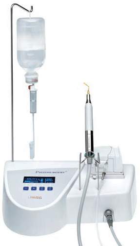
Piezoelectric surgical unit
It features a foot switch and a piezoelectric hand piece that are linked to a main unit that provides electricity and a peristaltic pump for watering. An interactive key board operates the device. The two programs are called Root and Bone. Depending on the bone's quality, the power in the Bone program can be adjusted to any of the four levels. Perio or Endo power settings are available in the Root software. [[Figure 1]]
The handpiece has several inserts available in different kits
Basic Kit
Uses
Corticotomy
Ostotomy
Osteoplasty
Ostectomy
Equipped with
1 insert OT7,1 insert OT2,1 insert EX1,1 insert OP1,1 insert OP3
Sharp insert tips
The bone structures can be treated gently and effectively thanks to the insert tips' sharp edge. When a precise and well-defined cut in the bone structure is needed during an osteotomy, sharp insert tips are employed. Additionally, there are insert tips with sharp edges that are employed in bone chip harvesting and osteoplasty procedures.
The diamond-surfaced smoothing insert points allow for regulated and exact work on the bone structures. Smoothing insert tips are utilized in osteotomy when complex and delicate structures need to be prepared. For instance a sinus window or nerve access. Smoothing insert tips are used in osteotomy to achieve the desired final bone shape.
Blunt insert tips
Blunt insert tips are used for preparing the soft tissue. For example, for elevating Schneider’s Membrane or for lateralizing nerves. In periodontology, these tips are used for root planing.
Insert tips color
Gold For all insert tips used to treat bone. A coating of titanium nitride is applied to obtain the gold color, it also improve the surface hardness which means a longer working life.
Steel For all insert tips used to treat soft tissue or delicate surfaces such as the roots of teeth.
The sound of the cutting can be utilized as an auditory feedback to gauge the force applied. Piezosurgery produces a very precise cut with very little force required. When too much pressure is exerted, the tip becomes immobile and produces heat. The harvesting methods, ridge splitting, and sinus lift operations that can be performed with comfort and convenience employing piezosurgery are of special interest to implantologists.
Harvesting bone chips
In addition to supporting growth factors at the recipient site to hasten bone repair, bone chips play the roles of creating space and acting as guides for bone regeneration (via osteoconduction).
The gold standard in periodontology is autogenous bone grafts, which are indicated in specific cases. Greater healing prospects are offered by autologous bone in large flat defects surrounding teeth. Because of the anatomical conditions, the success probability of regeneration measures in particular abnormalities is low. There are benefits to using bone chips to treat the deformity. Using the osteoplasty No. 3 instrument, the bone can be extracted from the linea obliqua and reinserted into the defect.
Bone blocks
Particulate grafting material utilized in nonstable environments may have limited effects on bone repair. Particulate material is effective in areas surrounding by bone, particularly when membranes are employed. But in methods involving either vertical or horizontal augmentation, the particle materials exhibit their limits. Bone blocks work best in these situations. The chin, linea obliqua, and crista iliaca are traditional donor sites. Utilizing piezosurgery facilitates the linea oblique technique. The adjacent structures are protected from harm by the instrument tip's low amplitude, the best cooling effect, and the selective cut. No direct view of the deep horizontal cut, no preparation of the N. mentalis is required, and a modest access that permits undermining preparations is adequate.
Bone splitting
The downside of the older methods is that they usually required membranes and a second surgical (donor) location. Furthermore, a second procedure is typically required once particle or bone block grafts are used, as implants cannot be placed concurrently. Although particle matter and implant implantation can coexist, the outcome may be jeopardized if the graft material micromoves. When there is enough bone height but not enough width, bone splitting can be necessary. Numerous difficulties are avoided, including the necessity for membranes and the utilization of biomaterials or transplants. An enhanced split thickness flap prevents bone resorption. The buccal and palatal plates are separated by the bone-splitting process, respectively. The resulting gap between the two plates is perfect for implant integration and regeneration. Bone envelops augmentation materials, which have bidirectional blood vascular supply and cell migration. No micromovement is present. These are the perfect circumstances for low-risk recovery. However, pressure trauma poses a danger of bone splitting, particularly in the D1 bone. Because the periosteum is not raised, fractures do not pose a threat. Usually, this greenstick fracture mends without any issues. Since bone flexibility is higher in the maxilla, bone splitting has been applied there most often. In densely mineralized bone, piezosurgery is utilized because the width of the bone prevents implant implantation while the vertical dimension of the bone is preserved. To complete the case in one stage, bone splitting is done.55 It is necessary to cover the bone with a split thickness flap because the alveolar crest will enlarge after splitting. In these circumstances, a tension-free suture is feasible. With its blood vessels, the periosteum remains attached to the bone. A saw-shaped tip is used to make an incision (osteoplasty No. 5). There is no need for a buccal side releasing incision because the bone is elastic. Osteotomes can be used to lengthen the bone, and a combined drill-split method can be used to introduce implants. The gap between the two lamellae can be filled in using the bone chips that are remaining from the drills. It is possible to perform a tension-free suture without using membranes.
Sinus floor elevation
For the treatment of vertical deficits in the posterior maxilla, the sinus floor elevation is currently a standard procedure. Most often, lateral access is used. The process of creating the schneiderian membrane involves modifying the Caldwell-Luc method. With this process, there is a chance of membrane perforation either during the window preparation phase or during the elevation stage. Membrane perforation is a potential in circumstances where sinus-mouth communications have recently closed, either due to septae presence or for other causes. This puncture frequently develops into a rupture that prevents closure, even using membranes or microsutures. A intact membrane is necessary to keep the transplant stable. Piezosurgery lowers a number of the hazards connected to sinus lift operations. It is nearly hard for the clinician to damage the membrane when preparing the window because of the selective cut made by the instrument. The osteoplasty No. 5 tip is recommended in instances with a thin bone wall, while the osteoporoplasty No. 1 tip is utilized for reduction in cases with thick bone before the osteoplasty No. 5 tip is employed. For the transplant process, the leftover bone chips are gathered. The membrane can be raised 2 mm across the window's perimeter using manual equipment, which are utilized for the membrane preparation. The Piezosurgery EL2 and EL3 elevating tools are used following this procedure. With hydropneumatic pressure delivered through the cooling saline solution and micro-saw settings, these instruments function like traditional sinus instruments.
The risk of membrane rupture during sinus lift is decreased from 30% to 70% when piezosurgery is used. [12], [13]
Application of ultrasound in osteoconduction
Ultrasound therapy has developed within the field of physiotherapy since its initial application in 1932, mostly for the treatment of soft tissue problems. The 1990s saw the publication of the first prospective, randomized, double-blind clinical trials. According to the findings of these investigations, applying ultrasound could shorten the healing period of newly fractured tibia and radial bones by 38%. Here, a transducer was applied to the skin across the fracture, and low intensity pulsed ultrasound was used for 20 minutes every day at an intensity of 30 mW/cm2. It also turned out that ultrasonography might be used to treat slow-moving or nonuniting fractures. Moreover, it appeared that the effects of ultrasound might promote bone healing following osteotomy or osteodistraction in addition to fracture repair. Thus, it was determined that ultrasonography might have the ability to promote maxillofacial bone repair. It would seem logical to believe, based on the research, that ultrasound affects bone cells throughout the healing process; nevertheless, a reported effect might be attributed to the mechanical and circulatory conditions at the location. Furthermore, there is no proof to support the claim that low-intensity pulsed ultrasound (LIPUS) induces osteoconduction in a rat mandibular bone defect wrapped in a collagen membrane [Schortinghuis J, 2005]. [14]
Ultrasound may help to regrow teeth?
According to studies, high-intensity focused ultrasound can noninvasively stop blood vessel bleeding; on the other hand, LIPUS can effectively release preformed fibroblast growth factors from a macrophage-like cell line and promote angiogenesis during wound healing. improves bone development into titanium porous implants, accelerates resorption healing with reparative cementum, improves mandibular growth in developing baboons, and improves bone healing following fractures and mandibular osteodistraction. For recurrent aphthous stomatitis, routine use of low-intensity ultrasound seems to have a modestly positive impact. The prototype ultrasonic gadget can be installed on a removable plastic crown or on braces. The tooth root can be stimulated to grow by gently massaging the gums with a wireless device that is about half the size of a newborn fingernail. Although LIPUS appears to be useful in healing and restoration, more research is needed to understand how they contribute to periodontal regeneration. Studies that specifically address LIPUS's function in periodontal regeneration are still lacking. [12], [13], [15], [16], [17], [18]
Conclusion
Using ultrasonography to analyze periodontal structures in three dimensions is one way that mega Hertz ultrasound is used for diagnostic purposes. The benefit of employing noninvasive evaluations devoid of ionizing radiation may find uses in clinical assessment and treatment planning before implant implantation, as well as in the assessment of soft and hard tissue healing following periodontal surgery (regenerative, mucogingival, or implant surgery). In the field of medicine, ultrasonic equipment has been shown to be successful and efficient in treating periodontal disease. When used correctly, ultrasonography is less taxing on the operator, takes less time to recover, and is gentler on soft tissues. Although LIPUS appears to be useful in healing and restoration, more research is needed to understand how they contribute to periodontal regeneration. A relatively recent surgical method for periodontology and implantology called piezosurgery can sometimes be utilized in place of or in addition to more conventional oral surgical procedures. A precise cut is made possible by the instrument's low pressure, and the selective cut also typically protects delicate tissues.
Source of Funding
None.
Conflict of Interest
None.
References
- WR Laird, AD Walmsley. Ultrasound in dentistry. Part 1--Biophysical interactions. J Dent 1991. [Google Scholar]
- G.F.S. The physical and biological effects of high-frequency sound-waves of great intensity. J Franklin Inst 1927. [Google Scholar]
- MC Catuna. Sonic Energy; A possible dental application. Preliminary report of an ultrasonic cutting method. Ann Dent 1953. [Google Scholar]
- DD Zinner. Recent ultrasonic dental studies, including periodontia, without the use of an abrasive. J Dent Res 1955. [Google Scholar]
- WN Johnson, JR Wilson. The application of ultrasonic dentalunits to scaling procedures. J Periodontol 1957. [Google Scholar]
- WR Laird, AD Walmsley. Ultrasound in dentistry: Part 1 biophysical interactions. J Dent 1991. [Google Scholar]
- AM Pattison, L Pattison, MG Newman, HH Takei, PR Klokevold, FA Carranza. Scaling and root planning. Carranza’s Clinical Periodontology 2006. [Google Scholar]
- N Frederiksen, SC White, MJ Pharoah. Oral Radiology; Principles and Interpretation. Oral Radiology; Principles and Interpretation 2004. [Google Scholar]
- AR Williams. . Ultrasound: Biological Effects and Potential Hazards 1983. [Google Scholar]
- B S Khambay, A D Walmsley. Acoustic microstreaming: detection and measurement around ultrasonic scalers. J Periodontol 1999. [Google Scholar]
- SJ Ewen. Ultrasound and Periodontics. J Periodontol 1960. [Google Scholar]
- G Messiner, B Oehme, J Strackeljan, A Kuhr, T Kocher. A method for the validation of a calculus detection system. J Clin Periodontol 2005. [Google Scholar]
- VK Bains, R Mohan, R Bains. Application of ultrasound in periodontics: Part II. J Indian Soc Periodontol 2008. [Google Scholar]
- J Schortinghuis, ALJJ Bronckers, B Stegenga, GM Raghoebar, LGM de Bont a. Ultrasound to stimulate early bone formation in a distraction gap: a double blind randomised clinical pilot trial in the edentulous mandible. Arch Oral Biol 2005. [Google Scholar]
- VO Kasat, H Saluja, BM Rudagi, JV Kalburge, S Sachdeva. Malignant fibrous histiocytoma of maxillary alveolar ridge extending to the hard palate. J Cancer Res Ther 2014. [Google Scholar]
- HM Saluja, S Sachdeva, A Mani. Role of reactive oxygen species and antioxidants in periodontal disease. J Cell Biotechnol 2021. [Google Scholar]
- SN Shah, MV Awinashe, A Mohammed, D Agarwal, A Anand, TP Sonkar. Antimicrobial Efficacy of Sodium Hypochlorite, Neem, Tulsi, and Aloe Vera as a Root Canal Irrigants Against E. Faecali. Int J Health Sci 2022. [Google Scholar]
- H Saluja, S Sachdeva, S Shah, A Dadhich, P Tandon, V More. Autogenous Grafts for Orbital Floor Reconstruction: A review. Int J Oral Craniofac Sci 2017. [Google Scholar]
- Introduction
- Discussion
- Application of ultrasound in diagnosis
- Application of ultrasound imaging for periodontal assessment
- Application of ultrasound in calculus detection
- Application of ultrasound in scaling and root planing
- The mechanism of removing calculus
- Mechanism of removing biofilm or plaque
- Endotoxin removal and root detoxification
- Application of ultrasound in curettage
- Application of ultrasound as microultrsonics
- Application of ultrasound as ultrasonic cleaner
- Application of ultrasound in bone surgery
- Indications
- Advantages
- Disadvantages
- Piezoelectric surgical unit
- The handpiece has several inserts available in different kits
- Basic Kit
- Equipped with
- Sharp insert tips
- Blunt insert tips
- Insert tips color
- Harvesting bone chips
- Bone blocks
- Bone splitting
- Sinus floor elevation
- Application of ultrasound in osteoconduction
- Ultrasound may help to regrow teeth?
- Conclusion
- Source of Funding
- Conflict of Interest