Introduction
The loss of teeth due to trauma, periodontal disease, or extraction is one of the main causes of bone volume loss for implant placement in the anterior tooth region. Additionally, the labial alveolar bone frequently experiences rapid resorption following natural tooth loss, with a volume decline of around 25% during the first year and a width decrease of 40–60% over the subsequent three years. In the aesthetic zone, such a flaw management becomes crucial.
In order to enlarge horizontal ridges while retaining the periosteal connection, ridge split or ridge expansion was first used in the early 1970s. This procedure involved meticulously enlarging the cortical plates. The additional benefit of this method was that it allowed for both implant placement and augmentation in a single visit. For handling narrow edentulous ridges (>3.5 mm) for implant insertion, ridge-splitting procedures are more effective in the maxilla than in the mandible. Therefore, less invasive procedures like ridge splitting and expansion can be easily performed on patients with moderate horizontal ridge abnormalities without causing too much damage.
This paper describes a case of trauma-related loss of the upper central incisors. Using the ridge split approach, the patient was given an implant-supported prosthesis for rehabilitation.
Case Report
The main complaint of a 40-year-old male patient who visited the Division of Prosthodontics was that his top front teeth were missing. The patient, who was wearing a removable partial denture at the time, revealed that he had lost 11 and 21 in an accident two years back. Kennedy's Class IV edentulous space with respect to 11, 21, and Sibert's Class I defect in the 21 region were discovered during an intraoral examination.
The patient's medical history revealed that he was average in built and fit to undergo the surgery. A thorough case history was completed. The patient underwent various preoperative tests and procedures such as a traditional orthopantamogram, a cast for ridge mapping, oral prophylaxis, and standard blood and urine tests. It was discovered using ridge mapping that the bone in region 21 contains a Seiberts Class I flaw.
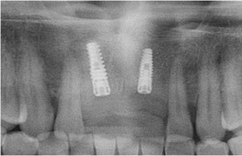
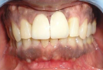
The treatment strategy was developed. A 3.8 x 13-mm implant was inserted in area 11. A ridge split was performed with regard to 21 region, and a 3.3 x 11 mm implant was inserted. The second part of surgery was carried out after a six-month recuperation period. PFM crown was given for the patient's rehabilitation.1, 2
Surgical procedure
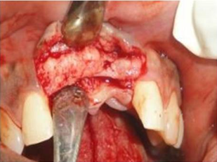
7.5% w/v Povidone-Iodine solution was used to clean the surgical site. 2% lignocaine combined with 1:80,000 epinephrine was used to numb the area. By making a precise mid-crestal incision and then raising a full thickness flap that measured about 3-5 mm bucco-palatally in area 21, the ridge crest was exposed [Figure 1]. A 15-number blade was used to make a mesiodistal incision, leaving 1 millimeter of bone intact on either side. Sequential osteotomies for expansion were performed using a micro-saw handpiece and SPLITMASTER II.
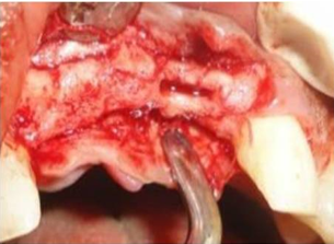
In order to stretch the base of the bone into a V shape, the initial length of the osteotome was created 3 mm deeper than the required implant length of 13 mm [Figure 2]. This was done by inserting progressively larger diameter osteotomes that were 0.5 mm shorter than the preceding instrument. Because each successive osteotome was inserted and removed delicately, maintaining the bone's resilience, the visco-elastic property of the bone was used to prevent fracture.
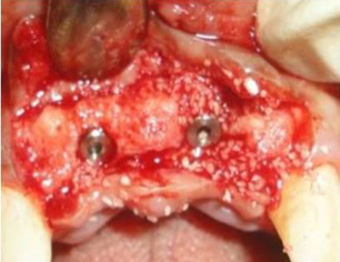
Two equinox implants measuring 3.8 mm by 13 mm [Figure 3] were inserted, and after putting the cover screw, demineralized freeze-dried bone allograft was used to fill the area between the implants. In order to establish tension-free interrupted sutures that result in close proximity, a coronal flap was raised over the implant by making a periosteal-releasing incision.
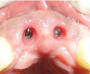
The patient received both verbal and written postoperative instructions after the placement of the periodontal pack. Analgesics, antibiotics, and chlorhexidine mouthwash 0.2% were recommended for five days. The periodontal pack and stitches were removed seven days after surgery. The patient underwent prosthetic rehabilitation with full PFM crowns after being periodically evaluated for the next 8 months. [Figure 4, Figure 5, Figure 6].
Discussion
As only 4-6 mm of bucco-palatal bone, width is available with/without a "hourglass" facial deformity, alveolar ridge augmentation is necessary for implant treatment plans in deficient ridges. This helps to reduce the stress at the crestal bone region.,3, 4 Therefore, ridge augmentation before implant insertion is frequently necessary for long-term prosthesis durability. Positive results are only anticipated if there is at least a 6 mm ridge width available, resulting in at least 1-1.5 mm of bone surrounding the implant.5
Dr. Hilt Tatum developed a method of ridge-splitting bones in the 1970s. He specifically used tapered channel formers and D-shaped graduated osteotomes/wedges in this technique. Before 1985, he placed more than 5000 maxillary anterior implants utilizing the ridge splitting technique, which allowed him to enlarge atrophic ridges that were less than 3 mm long while leaving the periosteum unharmed.6 Summers and Scipioni et al. revived the edentulous ridge expansion procedure in 1994 and published several articles on it. The approach has a 98.8% implant survival rate for more than 5 years.6
The ridge split technique has become a mainstay of implant dentistry with the development of microsaws, piezosaws, and other contemporary implant dentistry procedures. Previously, bone expansion treatments were only recommended in areas with high bone density and division B bone volume. But now, because of the dynamic viscoelastic nature of the bone, even thinner ridges (3.5 mm) can be expanded with better-controlled instrumentation with reduced danger of fracture, trauma, and bone. The softer the trabecular bone quality, the lower the elastic modulus and the greater the viscoelastic nature of the ridge. Therefore, it is anticipated that the less dense bone will expand predictably and more easily.7, 8
Bone expansion gives a more normal facial contour to the region. The bigger palatal bone is more difficult to manipulate, therefore bone splitting does not influence the facial and palatal plates equally. As a result, the expansion process occurs predominantly in the direction of the thinner facial plate. Osteotomes are used to further expand the osteotomy site using controlled sequential mild tapping (approximately 1 mm for every tap) with a surgical mallet. The osteotomy is first prepared 2-4 mm deeper than the eventual implant length using an initial 2 mm drill.
Before using an osteotome to prepare the site and when inserting the implant, it is important to feel the labial tissue.6, 7 The golden rule to follow is that if there is any uncertainty about a perforation, the labial tissue should be reflected, and the site should be examined. If a perforation does occur, an autogenous graft and/or barrier membrane are then employed to augment the site. A physio-dispenser handpiece with a moderate speed and high torque is used to thread the final implant into place. In order to reduce the danger of crestal bone loss and to help with bone remodeling, bone graft can be put between the bone and the implant and at the crestal region with membrane.9
Conclusion
Many techniques for the placement of implants in severed alveolar ridges have been established. Especially where ridge split or spreading needs to be advocated with a ridge width less than 3.5 mm. Careful patient selection is the mainstay in the success of ridge split technique which requires bone elevation. Most of the time, these splited alveolar ridges heals as the bone fracture does, with a clot filling the increased gap over time before being replaced by woven bone and then, at the interphase, by load-bearing lamellar bone. The surgical technique utilized in this case can be applied to either jaw, but the maxilla is the superior candidate. In this case report, the satisfactory treatment outcomes of implant dentistry where augmentation of severed alveolar ridges is an important aspect have been highlighted in detail.

