Introduction
The frenum is anatomic structure derived from the latin word “fraenum”, which is formed by a membranous fold of mucous membrane, connective tissue and sometimes muscle fibres. There are numerous frena which are present in the oral cavity and the most commonly noted frena are the maxillary labial frenum, the mandibular labial frenum, and the lingual frenum.1
The stability to the upper and lower lips and to the tongue is provided by the frena which is its primary function.2
The aberrant frenum can lead to gingival recession when they are too closely attached to the gingival margin. This can be either because of opening of the gingival crevice due to muscle pull or a interference with the improper placement of a toothbrush.1
The development of maxillary labial frenum is from the post-eruptive remnant of the ectolabial bands which connects the tubercle of the upper lip to the palatine papilla. There is no bone deposited inferior to the frenum when the two central incisors erupt widely separated. A V-shaped bony cleft is formed between the two central incisors, resulting in an abnormal frenum attachment. When there is a decreased vestibular depth and an inadequate width of the attached gingiva, mandibular frenum is considered as aberrant.1, 3 Frenum has been classified by Placek et al (1974) depending upon the extension of the attachment fibers as:4
Mucosal – here the frenal fibers are attached to the mucogingival junction
Gingival – here the fibers are inserted within the attached gingiva
Papillary – here the fibers are extending into the interdental papilla
Papilla penetrating – here the frenal fibers cross the alveolar process and extend up to the palatine papilla.
Papillary and papilla penetrating frena are considered as pathological and have been associated with recession, loss of papilla, diastema, malalignment of teeth, difficulty in brushing and it may also prejudice the denture fit or retention leading to psychological disturbances to the individual.5, 6 Miller has recommended that the frenum should be characterized as pathogenic when the frenum is unusually wide or there is no zone of attached gingiva along the midline or when the frenum is extended the interdental papilla shifts.7
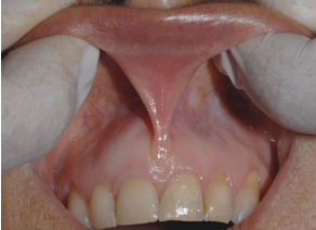
The abnormal frena can be diagnosed visually by moving the upper lip outwards and downwards and lower lip is moved outwards and upwards. If the gingival margin shows movement or if blanching is seen due to ischemia, then the test is positive and the frenum is said to be aberrant This test is known as the Blanching test or Tension test.(Figure 1)
According to Olivi et al, clinical indications for frenum removal include: 8
Abnormal frenum with inflamed gingiva due to poor oral hygiene
Abnormal frenum associated with inflamed gingival recession
Maxillary frenum associated with Diastema after complete eruption of permanent canines
Abnormal maxillary frenum (Class III or IV), resulting in the presence of a diastema during mixed dentition.
Abnormal mandibular frenum with high insertion leads to the onset of gingival recession
The treatment of the aberrant frena can be either done by frenectomy or by frenotomy procedures. Frenectomy is defined as the complete removal of the frenum, with its attachment to the underlying bone, while Frenotomy is the relocation of the frenal attachment.9
There are various surgical techniques of frenectomy which include Classical frenectomy by Archer and Kruger, Millers technique (unilateral single pedicle flap), Schuchardt Z-plasty, V-Y Plasty, Frenectomy using electrocautery, Laser – Diode,CO2, Nd:YAG, Er:YAG and other soft tissue lasers.
The main drawback of the classical frenectomy technique is scarring which may lead to periodontal problems and unesthetic appearance.
Miller has given a surgical technique where the frenectomy is combined with a laterally positioned pedicle graft. The primary advantage of this technique is complete closure seen across the midline due to laterally positioned gingiva and the healing by primary intention which resulted in aesthetically acceptable attached gingiva across the midline. Interdental papilla remained undisturbed as no attempt was made to dissect the trans-septal fibers. Therefore esthetically and functionally better results were obtained.7
This article presents a case report on frenectomy by Millers technique.
Case
A 23 years old male patient reported to the Department of Periodontology. On clinical examination, according to Miller, the frenum was considered abnormal as it was unusually wide and there was inadequate zone of attached gingiva along the midline7 and also the tension test was positive and therefore frenectomy by Millers technique was decided. The entire surgical procedure was explained to the patient and written informed consent was obtained from the patient before the surgical procedure.
Surgical technique
Armamentarium - no.15 surgical blade, Haemostat, Gauze, 5-0 black silk sutures, Needle holder, suture cutting scissors and a periodontal dressing.
2% lignocaine with 1:80000 adrenaline was used as an local infiltration to anaesthetized the area. To separate the frenulum from the interdental papilla a horizontal incision was made. This incision was extended apically up to the vestibular depth to completely separate the frenum from the alveolar mucosa. (Any remnant of frenum tissue in the mid line and on the under surface of lip was excised). 2-3 mm apical to marginal gingiva up to vestibular depth ,an vertical parallel incision was made on the mesial side of lateral incisor The gingiva and alveolar mucosa in between these two incisions were undermined by partial dissection to raise the flap. 1-2 mm apical to gingival sulcus in the attached gingiva a horizontal incision was made, connecting the coronal ends of the two vertical incisions. Flap was raised, mobilised mesially and sutured to obtain primary closure across the midline. Postoperative instructions were given to the patient. Patient was recalled 1 week postoperatively for the removal of the sutures.
Discussion
To create more functional and aesthetic results more conservative and precise techniques are being adopted in this era of periodontal plastic surgery, 10 Management of abnormal frenal attachment started from Archers classical frenectomy to modern concepts by Edwards. To evade the formation of scar and to facilitate healing, application of laser and soft tissue grafts helped in evolving the newer frenectomy procedures.11 The features that help in assessment of a frenum are vestibular depth, attached gingival zone, interdental papilla and midline diastema. An adequate zone of attach gingival gives an aesthetically pleasing appearance and also help in the avoidance of recession which necessitates removal of the same.11
Frenum is said to be pathogenic when it is unusually wide or there is no apparent zone of attached gingiva along the midline or the interdental papilla shifts when the frenum is extended.7
In a study conducted by Miller on 27 subjects with abnormal frenum who had undergone orthodontic closer of diastema, frenectomy combined with a laterally positioned pedicle graft was performed. The study showed that there was no loss of interdental papilla. In 24 cases there was no relapse of diastema was seen and in only three cases minimal relapse (less than 1 mm) was noted. He suggested that the reopening of diastema was prevented by the newly formed broad attached gingiva which contains collagenous fibres which may have a bracing effect. According to Miller theideal time for performing this surgery must be after orthodontic movement is completed and about six weeks before appliances are removed.7
The Millers technique offers two distinct advantages. Firstly, on healing, there is no anesthetic scar formation as a continuous band of gingiva ids formed across the midline and secondly there is no trauma to the interdental papilla as the trans-septal fibres are not disrupted surgically.
In a study done by Nirwal Anubh et al,12 performed frenectomy using laterally displaced pedicle graft. They achieved esthetically pleasing result as there was no loss of interdental papilla and no scar formation in the midline. Similar results were obtained in the present case report with good colour match.
A similar study was conducted by Ameet Mani et al13 and Devishree et al14 where they performed lateral pedicle frenectomy. They too observed that healing was by primary intention without any scaring in the midline.
In our case report, postoperatively we were able to achieve esthetically pleasing result without scar formation. Assessment of Pain was done using the Visual analogue scale during and after 24and 72 hrs. Mild to moderate pain was there during and soon after the procedure. Post operative review after 2 days reveals absence of pain in the surgical site.15
Conclusion
The present case report describes the Millers surgical technique i.e., frenectomy with a lateral pedicle graft. The main advantage of this techniques is that the healing takes place by primary intention, a thick zone of attached gingiva is formed, the colour matches with the adjacent tissue, no scarring takes place and there is no loss of interdental papilla as the transseptal fibers are not severed. We were able to achieve the above in our Case. More Comparative studies need to be done.


